 ResourceYear 2HistologyBy: ASU Faculty Staff
Trachea to alveoli — histology comparison chart
Loading document preview...
Please wait while the PDF stream connects
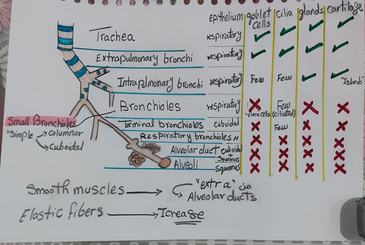
Comparison chart of epithelium, goblet cells, cilia, glands and cartilage from trachea to alveoli. Follow each airway segment across the columns. The drawing also compares smooth muscle and elastic fibres. The caption attributes the original drawing to department teaching staff.
Having trouble reading?Open directly in Google Drive
Use the fullscreen icon or zoom controls for distraction-free readingRelated Study Materials in this Module
View module →
 Playlist
Conducting and respiratory portions: NAS recordings — ASU2Learn
HistologyBy: ASU Faculty Staff